The ultrasound-guided distal sciatic nerve block is the ideal block for patients with distal leg and ankle injuries (large lateral leg laceration, pain reduction from bimalleolar fractures, Achilles tendon rupture, lower leg burns, abscesses, etc.). Depending on the anesthetic used, the block can facilitate fracture reduction or abscess drainage or be used as an adjunct in a multimodal plan for pain control. Unfortunately, the superficial structures of the medial lower leg and ankle are not innervated by the distal sciatic nerve, and a saphenous nerve block (distal aspect of the femoral nerve) may be needed if a more complete analgesia to the lower leg is desired.
Explore This Issue
ACEP Now: Vol 34 – No 06 – June 2015The block can facilitate fracture reduction or abscess drainage or be used as an adjunct in a multimodal plan for pain control.
Indications
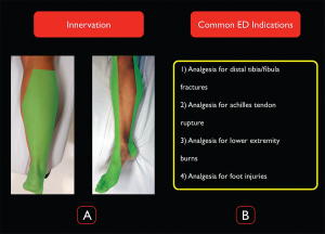
The distal sciatic nerve innervates the majority of the lower extremity below the knee, making it an ideal block for ankle and distal tibial/fibular fractures and injuries to the foot (see Figure 1). It does not provide anesthesia to the medial aspect of the lower leg, which is innervated by the saphenous nerve (a distal branch of the femoral nerve).

(Click for larger image)
Figure 1. Distal sciatic nerve innervation and common ED indications for the block. Credit: Arun Nagdev
Contraindications
Any injury that could potentially result in compartment syndrome is a relative contraindication to performing a distal sciatic nerve block. High-energy injuries such as a solitary tibial or mid-shaft tibial/fibular fracture are known to have high rates of developing compartment syndrome. Additionally, crush injuries or any injury with associated vascular

(Click for larger image)
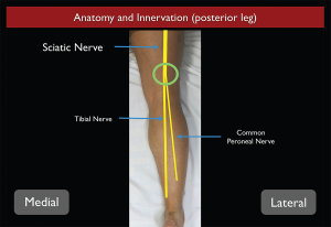
Figure 2. Sciatic nerve anatomy from the posterior aspect as it travels down the thigh into the leg. Credit: Arun Nagdev
compromise should trigger a detailed discussion with consultative services (orthopedics, trauma surgery, etc.) before an ultrasound-guided distal sciatic nerve block in the popliteal fossa is performed.
Anatomy
The distal sciatic nerve originates from the L4-S3 nerve roots of the lumbar-sacral plexus. The sciatic nerve initially runs deep in the posterior thigh, gradually becoming more superficial as it approaches the popliteal fossa. The large sciatic nerve bifurcates into the tibial (medial) and the common peroneal (lateral) nerves approximately 7–10 cm proximal to the popliteal fossa (see Figure 2). At this level, the sciatic nerve is bound by the semimembranosus and semitendinosus muscles medially and the biceps femoris muscle laterally. Although the sciatic nerve can be blocked at any location along its course, its superficial position in the region of the popliteal fossa makes this distal location ideal.1,2
Patient Positioning
If possible, the patient should be in the prone position, allowing easy access to the popliteal fossa and posterior aspect of the patient’s lower extremity. In a patient who is unable to lie prone (cervical spine immobilization, etc.), the affected extremity must be elevated and supported with mild flexion of the knee. This is best achieved by propping the foot with blankets or pillows to allow the ultrasound probe to easily fit between the popliteal fossa and the patient’s bed. Either position will allow the clinician to block the sciatic nerve in the popliteal fossa, with the prone position being technically much less difficult.
Equipment/Probe Selection
A high-frequency linear probe should be used to image the distal sciatic nerve in the popliteal fossa (see Figure 3). We recommend a 20- or 22-gauge, 3.5-inch (9 cm) spinal needle to reach the distal sciatic nerve because of its depth in the popliteal fossa. In addition, 20 mL of local anesthetic will be needed.
Pages: 1 2 3 4 | Single Page


No Responses to “How to Perform Ultrasound-Guided Distal Sciatic Nerve Block in the Popliteal Fossa”