Myth #5: CT of the Abdomen and Pelvis Has No Role in Evaluating Ovarian Torsion

Explore This Issue
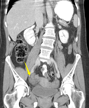
ACEP Now: Vol 39 – No 06 – June 2020Figure 2 (RIGHT)
CT depicting twisted follicle and enlarged ovary.
Patients with undifferentiated abdominal pain often undergo CT, but can CT assist in ruling in or out ovarian torsion? CT with IV contrast will often display findings suggestive of torsion.5,16,33,39–42 Findings on CT with high specificity for ovarian torsion include a twisted vascular pedicle (see Figure 2), a thickened fallopian tube with target/beak-like appearance, absent or reduced ovarian enhancement with contrast, and an enlarged ovary with a follicular ovarian stroma and peripherally displaced follicles.16,33,39–42 Features that are commonly found but not specific include an enlarged ovary, an adnexal mass, adnexal mass mural thickening, free pelvic fluid, fat stranding surrounding the ovary, uterine deviation toward the torsed ovary, and ovarian displacement toward the uterus.16,33,39–42 CT with contrast demonstrates a high sensitivity for these secondary findings, approaching 100 percent.16,33,39–42 If one of these secondary findings is present, TVUS and OB/GYN consultation should be expedited. If these findings are not present and the ovary is normal in size, TVUS may not be needed, depending on the any changes in the clinical course. If the ovary is abnormal on CT, then obtain TVUS.
Finally, if suspicious of torsion based on your history and exam, an ob-gyn consultation should be initiated prior to imaging. If an ob-gyn is unavailable, a general surgery consultation is warranted. Torsion is a time-sensitive condition; early involvement of specialists is paramount.
Key Point: A normal CT of the abdomen and pelvis with contrast that has no secondary findings displays high sensitivity for excluding ovarian torsion. If secondary findings such as an enlarged ovary are present, then obtain TVUS.
Case Conclusion
The CT of the abdomen and pelvis reveals a normal appendix but an enlarged right ovary. A small amount of pelvic free fluid and fat stranding around the right ovary are observed. You consult the ob-gyn on call, who requests a TVUS. The TVUS reveals an enlarged ovary with decreased venous flow on Doppler. The ob-gyn evaluates the patient and takes her to the operating room, where detorsion is successful.
Dr. Long is an emergency physician in the San Antonio Uniformed Services Health Education Consortium at Fort Sam Houston, Texas. Dr. Koyfman (@EMHighAK) is assistant professor of emergency medicine at UT Southwestern Medical Center and an attending physician at Parkland Memorial Hospital in Dallas. Dr. Gottlieb is associate professor, ultrasound division director, and ultrasound fellowship director in the department of emergency medicine at Rush University Medical Center in Chicago.
Pages: 1 2 3 4 5 | Single Page


One Response to “Dispelling 5 Ovarian Torsion Myths”
July 5, 2020
Jerry W. Jones, MD FACEP FAAEMExcellent article. Very concise and right to the point.