In Part 1 of our two-part series on marine envenomations, we reviewed some general information on wound care, antibiotic use, antivenom, and explored some species-specific management. In Part 2, we continue to review species-specific management.
Explore This Issue
ACEP Now: Vol 40 – No 08 – August 2021Phylum: Cnidaria
Class: Hydrozoa
Fire Corals (Millepora alcicornis)
Location: Worldwide (excluding Hawaii) in reefs and shallow waters
Appearance: White to yellow-green seaweed-like growths fixed to rocks and coral. They possess tentacles that extend upward and are roughly 2 m in length.
Pathophysiology and Symptoms: Contact with tentacles causes painful, urticarial lesions that may become hemorrhagic and ulcerate. Symptoms usually resolve within 90 minutes, but they can last up to 72 hours with skin hyperpigmentation that can last several weeks. Rarely, patients will present with mild systemic symptoms (eg, nausea, vomiting, myalgias, dyspnea, anxiety, abdominal pain, headaches, etc.).
Management: Pain is best managed with vinegar. Steroid creams and oral antihistamines can be used for mild urticaria. If severe, oral steroids may be warranted.
Class: Anthozoa
Sea Anemones
Location: Worldwide in deep and coastal waters, often attached to coral or rock
Appearance: Anemones vary in appearance. Most are a single polyp with a cylindrical body. Their mouths are surrounded by cnidocyte-containing tentacles.
Pathophysiology and Symptoms: Anemone venom contains multiple enzymes including cytolytic/hemolytic toxins, neurotoxins, cardiotoxins, and protease inhibitors, which cause symptoms ranging from erythema, pruritis, and blisters to fevers, chills, fatigue, myalgias, and syncope. Skin changes can become permanent in the form of hyper-/hypopigmentation and keloid formation.1
Management: Pain is managed with vinegar. Other symptoms are managed with supportive care.
Phylum: Echinodermata
Class: Echinoidea
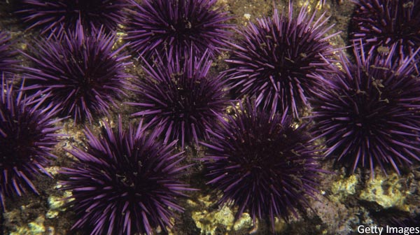
Sea Urchins
Location: Worldwide in both shallow and deep waters
Appearance: Composed of spherical, hard shells called “tests” that measure up to 4–5 inches in diameter which are covered in calcified spines. Venom is contained within these spines, as well as their pedicellarie (ie, pincers), which are more difficult to remove from human skin and contain more venom.
Pathophysiology and Symptoms: Contact causes an erythematous rash with localized burning, pruritis, myalgias (lasting approximately 24 hours), and edema. Symptoms rarely progress to nausea, vomiting, paresthesias, weakness, abdominal pain, hypotension, and syncope. Spines commonly break off, causing hyperpigmentation of the skin, and can lead to granuloma formation, secondary infection, and synovitis, if the joint is involved.
Management: Pain control is generally the biggest concern with these injuries and is best achieved with hot-water immersion and local lidocaine. Attempts to remove spines are often futile. The spines are very fragile and tend to crumble in the skin. To further complicate the removal process, areas where no spine remains may still have the appearance of a foreign body from “tattooing” of the skin. Operative exploration should be considered if there is joint involvement. Granulomas may also need surgical exploration because spines often crumble and are hard to find.
Pages: 1 2 3 4 5 | Single Page


No Responses to “Emergen-Sea Medicine Part 2: Overview of Marine Envenomations”