Explore This Issue
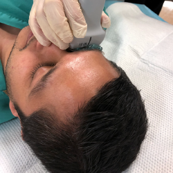
ACEP Now: Vol 38 – No 05 – May 2019Figure 3: Transverse visualization of the orbit.
Visualize the orbit in both transverse (see Figure 3) and longitudinal planes. After scanning through, the patient should be asked to move his or her eye right to left and up and down. A combination of still images and dynamic scanning clips will best document your exam.
Tips & Tricks

Figure 5: Stabilize your scanning hand by placing your pinky finger on the bridge of the patient’s nose.
Stabilize your scanning hand by placing your thumb or pinky finger (whichever is medial) on the bridge of the patient’s nose (see Figure 5). This will also prevent you from applying too much pressure
[/fullbar]
References
- Woo MY, Hecht N, Hurley B, et al. Test characteristics of point-of-care ultrasonography for the diagnosis of acute posterior ocular pathology. Can J Ophthalmol. 2016;51(5):336-341.
Pages: 1 2 | Single Page


No Responses to “High-Yield Ocular Ultrasound Applications in the ED, Part 3”