Optional
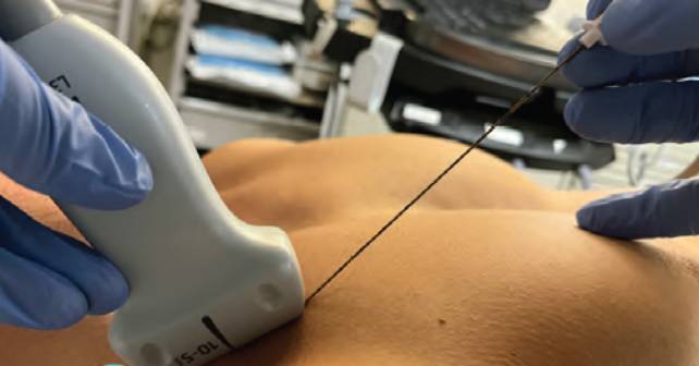
For simplicity and efficiency we recommend a multimodal pain strategy with a single injection site (either medial or lateral to the fracture site) and oral or intravenous medications. Clinicians who have additional time to perform the block can divide the anesthetic dosing and inject medial or lateral to the fracture site.8
Explore This Issue
ACEP Now: Vol 43 – No 02 – February 2024For all ultrasound-guided nerve blocks, we recommend placing documentation both in the EHR and as a small skin marking with a surgical pen on the patient. The cardiac monitor should be left on the patient for 30 minutes after the block to assess for local anesthetic systemic toxicity.
Conclusion
Clavicular fractures are common injuries in the ED. The CPB offers a novel technique that can be part of a multimodal pathway along with other agents (NSAIDs, acetaminophen, opioids, etc.) for the ED clinician. Our technique allows a rapid and simplified technique that can provide optimal pain control for patients with clavicle fractures.
Special thanks to our model Justin Bosley, MD.
Dr. Ashworth is a resident physician at Highland Hospital Alameda Health System in Oakland, Calif.
Dr. Lind is an attending physician at Highland Hospital Alameda Health System in Oakland, Calif.
Dr. Martin is director of the emergency ultrasound fellowship at Highland Hospital Alameda Health System in Oakland, Calif.
Dr. Nagdev is director for the emergency ultrasound division at Highland Hospital Alameda Health System in Oakland, Calif.
References
- Postacchini F, Gumina S, De Santis P, et al. Epidemiology of clavicle fractures. J Shoulder Elbow Surg. 2002;11:452-456.
- Kihlström C, Möller M, Lönn K, et al. Clavicle fractures: epidemiology, classification and treatment of 2 422 fractures in the Swedish Fracture Register; an observational study. BMC Musculoskelet Disord. 2017;18:82.
- Ashworth H, Martin D, Nagdev A, et al. Clavipectoral plane block performed in the emergency department for analgesia after clavicular fractures: A case series. Am J Emerg Med. 2023:S0735-6757(23)00539-9.
- Kukreja P, Davis CJ, MacBeth L, et al. Ultrasound-guided clavipectoral fascial plane block for surgery involving the clavicle: A case series. Cureus. 2020;12:e9072.
- Atalay YO, Mursel E, Ciftci B, et al. Clavipectoral fascia plane block for analgesia after clavicle surgery. Rev Esp Anestesiol Reanim (Engl Ed). 2019;66(10):562–563.
- Abu Sabaa MA, Elbadry AA, El Malla DA: Ultrasound-guided clavipectoral block for postoperative analgesia of clavicular surgery: A prospective randomized trial. Anesth Pain Med. 2022;12:e121267.
- Tran DQ, Tiyaprasertkul W, González AP: Analgesia for clavicular fracture and surgery: A call for evidence. Reg Anesth Pain Med. 2013;38:539-543.
- Lee CCM, Beh ZY, Lua CB, et al. Regional anesthetic and analgesic techniques for clavicle fractures and clavicle surgeries: Part 1-A scoping review. Healthcare (Basel). 2022;10:1487.
Pages: 1 2 3 4 | Single Page


One Response to “How To Perform an Ultrasound-Guided Clavipectoral Block”
February 28, 2024
Doc_CWhy not simply a superficial cervical plexus block more on the caudal side to get the rami claviculares of the cervical plexus?
Cheers