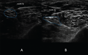
Figure 7A. In-plane lateral to medial approach of the needle. The needle tip is visualized clearly to ensure lack of nerve and vasculature puncture. Figure 7B. Spread of anechoic local anesthetic surrounding the distal sciatic nerve indicating a successful nerve block.
By Joseph Harrington
|
on June 10, 2015
|
0 Comment


No Responses to “Figure 7A. In-plane lateral to medial approach of the needle. The needle tip is visualized clearly to ensure lack of nerve and vasculature puncture. Figure 7B. Spread of anechoic local anesthetic surrounding the distal sciatic nerve indicating a successful nerve block.”