- Injuries to the to the medial upper arm are not covered by brachial plexus blocks.
- Interscalene blocks often provide better analgesia for shoulder dislocations.
- For injuries to the hand (distal to the wrist crease), a forearm nerve block may be more appropriate.
Anatomy
The brachial plexus is composed of nerves from the C5 to T1 nerve roots that travel from the cervical spine and innervate the neck, axilla, and arm. The brachial plexus branches as it travels from the cervical spine to the upper extremity, and the location of a block should be tailored to cover the nerve distribution of the injured region.
Explore This Issue
ACEP Now: Vol 42 – No 04 – April 2023When performed correctly, the supraclavicular nerve block will provide motor and sensory blockade to the radial, median, ulnar, musculocutaneous, and axillary nerve distributions of the arm. This leads to almost complete anesthetization of the arm, sparing the shoulder joint (suprascapular nerve) and the medial side of the upper arm (intercostobrachial nerves).
Equipment
Overall, the supraclavicular brachial plexus block requires similar equipment as other ultrasound guided nerve blocks:
- An ultrasound system with high-frequency 15-6 MHz or 10-5 MHz linear transducer set at the nerve (or soft tissue)
- A block needle: Preferably a 20- or 21-gauge, blunt-tipped, regional-block needle
- Appropriately sized syringe to draw local anesthetic
- Sterile saline flushes for hydrodissection
- Transparent adhesive ultrasound probe cover (Tegaderm, etc.)
- Antiseptic (e.g., alcohol, povidone/iodine, chlorhexidine)
- Sterile ultrasound gel
Procedure
Set-up
Place the patient on a cardiac monitor and ensure stable intravenous access. Position the patient supine in a 45-degree semi-upright position. Position the ultrasound system across from the affected extremity so that the patient’s exposed neck and the ultrasound screen are in the same line of sight. We recommend placing either a pillow or roll of blankets beneath the ipsilateral shoulder to improve block ergonomics (Figure 1A: picture of screen contralateral plus pillows to roll the patient). Set the ultrasound system to the “nerve” preset and cover the linear (high frequency) transducer with a transparent adherent dressing. We recommend using sterile surgical lubricant as a coupling agent if possible.
Also, always perform and document a neurovascular examination prior to performing your block.

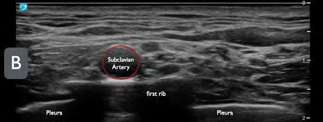
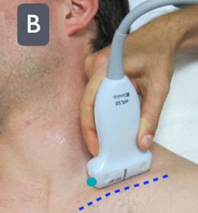
FIGURE 1B: Place the ultrasound probe (green dot indicating
probe marker) above the clavicle aiming the beam into the thorax. (Click to enlarge.)
Pages: 1 2 3 4 5 | Single Page


No Responses to “How To: The Ultrasound-Guided Supraclavicular Brachial Plexus Block”