
Explore This Issue
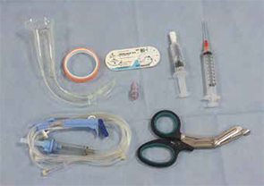
ACEP Now: Vol 34 – No 02 – February 2015Figure 1. Equipment used to make this laryngoscope tool.
The Case
A 42-year-old female presents to the emergency department after sustaining blunt facial trauma from a high-speed motor vehicle collision. She weighs more than 400 pounds and is back-boarded and collared. Blood is flowing freely from her mouth, and she is unresponsive to painful stimuli and gurgling with each respiration; this patient needs her airway secured five minutes ago. After the newly minted intern gives it a shot by direct laryngoscopy and pulls out with a look that says, “I think I just soiled my pants,” you step in. Using the GlideScope this time, you visualize the epiglottis, but then a splattering of
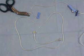
Figure 2. Cut the distal end of the IV tubing at an oblique angle.
blood hits the lens of your GlideScope and obscures the view. You try again with the same result. Is it time to dust off the #11 blade, or is there an alternative trick of the trade to improve visualization?
Background
In 1944, Bannister and Macbeth described direct laryngoscopy by aligning the pharyngeal, laryngeal, and oral axes to obtain direct visualization of the glottic inlet.1 Aligning these axes and visualizing the glottic inlet can be easily complicated by difficult airway characteristics (eg, large tongue, short neck, small mandible, bodily fluids in the airway, cervical immobility, facial trauma, edema, or limited mouth opening).2 Prior to the advent of video laryngoscopy, physicians had limited options to handle these complications before proceeding to a surgical airway. Video laryngoscopy can help circumvent these obstacles and has been shown to improve glottic visualization, especially in difficult airways.3 The hyperacute angle of the GlideScope can navigate challenging anatomy, and the video
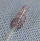
Figure 3. Fit the luer lock into the oblique IV tubing opening.
display provides a way to supervise novice operators. However, there is no solution for when the video laryngoscope’s view becomes obscured by bodily fluids. Removing the laryngoscope to clean it off wastes time and subjects the patient to another intubation attempt. To assist with clearing secretions, video laryngoscopes have been augmented using an inline suction device attached to the blade of the laryngoscope, but the results showed no improvement in time to intubation or increased success rates.4 Furthermore, the study did not address the complication of getting the secretions off of the screen to allow for better visualization.
To assist with clearing secretions, video laryngoscopes have been augmented using an inline suction device attached to the blade of the laryngoscope, but the results showed no improvement in time to intubation or increased success rates.4
Technique
Attaching IV tubing to a video laryngoscope can help improve visualization during
Pages: 1 2 3 | Single Page


One Response to “Keep Video Laryngoscope Clear with IV Tubing, Saline, and Some Ingenuity”
March 9, 2015
Kyle StevensWould love to see video of this DYI setup and of it in action