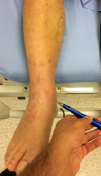
Case 2, Figure 3A: The deltoid ligament, below the medial malleolus.
Explore This Issue
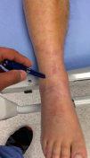
ACEP Now: Vol 37 – No 02 – February 2018Case 2, Figure 3B: Above the ankle joint, over the syndesmosis.
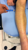
Case 2, Figure 3C: The proximal fibula.

Case 2, Figure 3A

Case 2, Figure 3B

Case 2, Figure 3C
In the clinic on day 10, X-rays were taken of the tibia and fibula (Case 2, Figures 4A and 4B). The proximal fibula shows the oblique fracture of the fibula. The fracture is only seen on the lateral view (Case 2, Figure 4B), a reminder of the orthopedic mantra, “One view is one view too few.” He had the classic Maisonneuve fracture, an external rotation mechanism resulting in injuries to the medial ankle (deltoid ligament or medial malleolus), syndesmosis, and interosseous membrane exiting out the proximal third of the fibula with an oblique fracture.
The patient was referred to an orthopedic surgeon and had surgery the following day, as shown in Case 2, Figure 2.
The OAR ask us to examine the posterior 6 cm of the medial and lateral malleoli, which was done. However, that is not a complete emergency department ankle exam. One needs to palpate many structures to localize the pain and discover the pathology. In this case, the ankle exam also revealed pain over both the deltoid ligament and the syndesmosis. Neither of these are part of the OAR. Either one can be a red flag on physical, and the combination certainly is concerning for a higher fibular fracture, which on palpation can be quite subtle. So in cases of twisting ankle injuries with significant medial ankle pain and little or no lateral ankle pain, it is best to X-ray the ankle and add the tibia/fibula views as a separate X-ray study.
This case again highlights that the OAR are not a complete emergency department ankle exam; they are simply a tool to use after a proper ankle assessment to decide if an X-ray is indicated.

Case 2, Figure 4A

Case 2, Figure 4B
It can be a bit daunting to see the “normal” X-rays and then the intraoperative films. This case was a missed Maisonneuve fracture, a classic fracture that will often show up on board certification examinations. More important than nailing the exam question, we should make sure we know how to recognize it the next time we see an emergency department patient with an ankle injury. This was a very subtle case without medial malleolus fracture (he had the deltoid ligament equivalent) and without shift of the ankle mortise. If we mistakenly put all of our focus on the X-ray, we are more likely to miss uncommon injuries. Again, the importance of history, appreciating that external rotation is a red flag, and physical tenderness over the deltoid ligament and the syndesmosis cannot be overemphasized. Then interpret the X-ray in the proper clinical context.
Case 3

Case 3, Figure 3


2 Responses to “Tips for Catching Commonly Missed Ankle Injuries”
March 3, 2018
abwExcellent article. Thank you.
July 30, 2018
Arun SayalThanks abw!
Happy to share these earls from our orthopedic surgeons – and from our patients.
Thanks to both groups for teaching me!
Arun