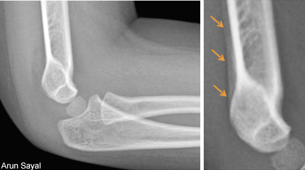
When assessing a patient with a suspected radiographically occult fracture, there are two options for the emergency physician: more tests or more time.
Explore This Issue
ACEP Now: Vol 39 – No 03 – March 2020More tests equates to additional X-ray views or advanced imaging (CT or MRI).
More time means treating the patient for the suspected diagnosis and arranging for a serial assessment.
I will discuss three cases and explore the ED management options.
Case 1: Occult Scaphoid Fracture
A 26-year-old female fell on an outstretched hand and has isolated wrist pain, tender snuff box, and scaphoid tubercle. X-rays of the wrist with scaphoid views are normal.
Diagnosis: suspected occult scaphoid fracture.
Follow-up studies have shown that 75 to 80 percent of patients with an ED diagnosis of a “suspected scaphoid fracture” do not have a fracture.1,2 There is concern that many patients are unnecessarily immobilized and require a low-yield follow-up appointment. These concerns have led some emergency departments to institute a wrist CT protocol during the initial visit in an attempt to definitively rule in or rule out a scaphoid fracture. A meta-analysis showed the sensitivity and specificity of CT for occult scaphoid fractures were 0.72 (95% CI, 0.36–0.92) and 0.99 (95% CI, 0.71–1.00), respectively.3 Even the CT may not definitively rule out a fracture and may be falsely reassuring. Additionally, if a patient’s radial-sided wrist pain comes from a partial scapholunate ligament (SLL) injury, the CT may be normal. If a patient subsequently falls during SLL healing (which may take weeks to months), the second force may convert a partial tear to a complete one, requiring operative management.
MRI is often considered the best advanced imaging option, as it shows the bone and soft tissues. A meta-analysis reported the sensitivity and specificity of MRI for occult scaphoid fractures were 0.88 (95% CI, 0.64–0.97) and 1.00 (95% CI, 0.38–1.00), respectively.3 Another smaller study showed early MRI missed 20 percent of radiographically occult scaphoid fractures.4 Therefore, normal MRI may not definitively rule out a fracture either. Additionally, high cost and low access prevent MRI from playing a role as an advanced imaging option for suspected occult scaphoid fractures during ED visits.
A bone scan may be considered due to a high sensitivity, though this modality is fading from common use. The sensitivity and specificity of bone scan for occult scaphoid fractures were 0.99 (95% CI, 0.69–1.00) and 0.86 (95% CI, 0.73–0.94), respectively, but there are many downsides to this imaging modality in the emergency department.3 For fracture detection, a bone scan generally requires 48 to 72 hours after injury to become reliably positive (though modern bone scans may need less time). Given its high sensitivity, a negative bone scan at 48 to 72 hours essentially rules out a fracture, but as with CT, a normal bone scan does not rule out a SLL tear. Unfortunately, a positive bone scan is hampered by low specificity. False positives can be generated by any condition that increases metabolic activity in bone, such as a bone contusion, infection, inflammation, degenerative joint disease, and tumors. Additionally, bone scans are associated with significant ionizing radiation (equivalent to 50 chest X-rays). Bone scans are fairly time-consuming and only available during certain working hours, and they require isotope availability. Bone scans miss important information including fracture pattern and/or precise location, making prognosis for that fracture difficult to assess. Therefore, a positive bone scan is often followed by a form of 3-D imaging (typically CT). As a result, radionuclide bone scans for suspected scaphoid fractures in the emergency department are largely impractical.


No Responses to “Tips for Managing Suspected Occult Fractures”