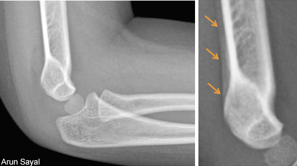
Suspected occult hip fractures, tibial plateau fractures, and cervical spine fractures, however, require immediate further evaluation, as they are more likely to displace if missed in the emergency department and not managed appropriately.9 These displacements can lead to more extensive surgery or surgery that may have been avoided altogether.9 In these cases, the need for advanced imaging during the index visit is evident.
Explore This Issue
ACEP Now: Vol 39 – No 03 – March 2020Patient Factors
Patient factors also play a role. Because of the tendency to displace with weight-bearing, patients with suspected tibial plateau fractures should be kept non-weight-bearing until confirmed or reassessed. For older patients, the strategy to immobilize, provide crutches, and require no weight-bearing can be a dangerous combination; fall risks are high. But younger patients may safely tolerate this approach, permitting immobilization and delayed advanced imaging in many instances. Patient factors around compliance and availability for follow-up should also influence our choice between more tests and more time.
Imaging Modalities
Advanced imaging for occult fractures in the emergency department generally refers to CT and MRI. Each has respective pros and cons.
A CT scan generally has high sensitivity for detecting fractures, and especially with 3-D reconstruction, it is an excellent tool for assessing bony alignment. CT provides little value for soft tissue injuries.
Musculoskeletal CT scans expose patients to ionizing radiation, but that exposure is far less than chest, abdomen, and pelvic protocols. A wrist CT is equivalent to the radiation of just 1.5–3 chest X-rays.10,11 A chest CT is equivalent to around 70; an abdomen/pelvis CT is equivalent to up to 100.12
MRI has advantages over CT. In addition to high sensitivity for fractures, MRIs can assess soft tissue structures—and without any radiation. However, high cost, long scan and radiology reading times, and poorer availability limit its role in the emergency department for occult fractures.
Bone scans and ultrasound in assessing suspected occult fractures are discussed above.
As a final consideration, the ED workup and treatment can vary from hospital to hospital based on local orthopedic preferences. Knowing how your local orthopedic surgeons prefer to manage the spectrum of suspected occult fractures from the outset optimally aligns initial ED care with the follow-up care patients will receive.
Summary
When considering advanced imaging, we are guided by the post-test probability for fracture; knowing the limits of plain films; understanding the complications of the suspected injury; the pros, cons, and indications for advanced imaging; and the proper ED treatment. Combining these helps optimize care.


No Responses to “Tips for Managing Suspected Occult Fractures”