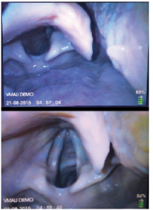
Figure 1 (Left). Blade positioning and Kovacs’ sign. In the upper image (ideal placement), the cricoid ring is not seen. There is more room beneath the posterior larynx on the monitor screen, which is critical for observing tube delivery. In the lower image, the cricoid ring and the internal aspect of the criothyroid membrane are visible between the vocal cords, indicating over-insertion of the hyperangulated blade and a steep angle of approach.
Figure 1 (Left). Blade positioning and Kovacs’ sign. In the upper image (ideal placement), the cricoid ring is not seen. There is more room beneath the posterior larynx on the monitor screen, which is critical for observing tube delivery. In the lower image, the cricoid ring and the internal aspect of the criothyroid membrane are visible between the vocal cords, indicating over-insertion of the hyperangulated blade and a steep angle of approach.
By Joseph Harrington
|
on December 15, 2015
|
0 Comment


No Responses to “Figure 1 (Left). Blade positioning and Kovacs’ sign. In the upper image (ideal placement), the cricoid ring is not seen. There is more room beneath the posterior larynx on the monitor screen, which is critical for observing tube delivery. In the lower image, the cricoid ring and the internal aspect of the criothyroid membrane are visible between the vocal cords, indicating over-insertion of the hyperangulated blade and a steep angle of approach.”