The Case
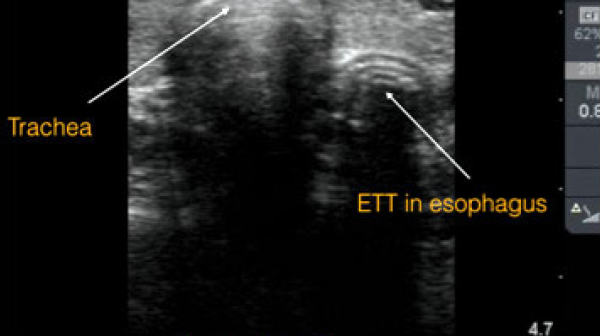
A 52-year-old patient is in cardiac arrest and needs endotracheal intubation. You’ve read some studies recently saying point-of-care ultrasound (POCUS) could confirm placement.
Explore This Issue
ACEP Now: Vol 38 – No 05 – May 2019Background
There are a number of ways to confirm endotracheal tube (ETT) placement. Quantitative waveform capnography is thought to be one of the best methods. However, in cardiac arrest, some studies suggest it is correct only about two-thirds of the time.1–3
An ACEP policy statement lists various methods to confirm ETT placement, which include:
- A physical exam (ie, auscultation of chest and epigastrium, chest wall movement, and condensation/fogging in the tube)
- Direct visualization or video laryngoscope of the tube passing through the vocal cords
- Pulse oximetry
- Chest X-ray
- Esophageal detector devices
- End-tidal carbon dioxide (CO2) detection (ie, continuous waveform capnography, colorimetric capnography, and non-waveform capnography)
Clinical Question
What is the accuracy of POCUS for ETT placement confirmation?
Reference
Gottlieb M, Holladay D, Peksa GD. Ultrasonography for the confirmation of endotracheal tube intubation: a systematic review and meta-analysis. Ann Emerg Med. 2018;72(6):627-636.
- Population: Prospective or randomized controlled trial (RCT) of adults undergoing assessment of transtracheal POCUS for ETT placement confirmation
- Excluded: Case reports, case series, retrospective studies, cadaver studies, pediatric studies, and conference abstracts
- Intervention: Transtracheal POCUS to confirm ETT placement
- Comparison: Confirmatory testing of ETT placement such as end-tidal capnography, colorimetric capnography, or direct visualization
- Outcome:
- Primary Outcome: Accuracy of transtracheal POCUS versus other forms of confirmation
- Secondary Outcome: Time to confirmation and subgroup analyses
Authors’ Conclusions
“Transtracheal sonography is rapid to perform, with an acceptable degree of sensitivity and specificity for the confirmation of endotracheal intubation. Ultrasonography is a valuable adjunct and should be considered when quantitative capnography is unavailable or unreliable.”
Key Results
The search identified 15 prospective observational studies and two RCT, including a total of 1,595 patients. A majority of the studies (12 out of 17) were performed in the emergency department. The mean patient age was 55 years, with 57 percent being male. The esophageal intubation rate was 15 percent.
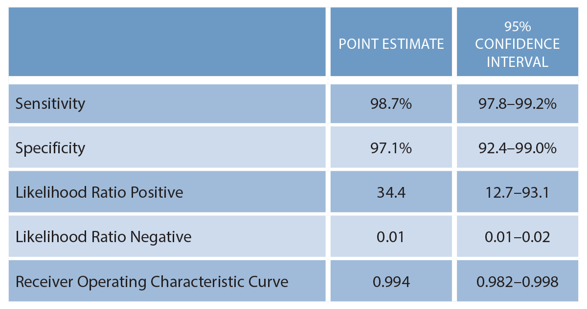
- Primary Outcome: Diagnostic accuracy of transtracheal POCUS for ETT placement (see Table 1)
- Secondary Outcome: Mean time to confirmation was 13.0 seconds (95% CI, 12.0–14.0).
- Subgroup Analyses: These did not demonstrate a significant difference by location, provider specialty, provider experience, transducer type, or technique.
Evidence-Based Medicine Commentary
- Included Studies: The 17 studies included were relatively small with wide confidence intervals. Fifteen of the 17 were observational studies. Only 216 patients (14 percent) were in RCTs. Thirteen of the 17 studies used convenience sampling instead of consecutive patients, which can introduce bias and thereby limit the strength of the conclusions.
- Lack of Gold Standard: A number of methods are available and often used in combination, but there is no gold standard. Each confirmation method has limitations. Auscultation can prove inaccurate, especially in loud environments. Chest X-ray takes too long, and as previously mentioned, capnography in cardiac arrest has low sensitivity.
- Esophageal Intubation Rate: This was very high (15 percent) and may be due to studies including medical students and residents in addition to attending physicians. A previous study has shown the rate of esophageal intubation in the emergency department to be only 3 percent.4
- Fast: POCUS for ETT placement was not only accurate but also fast, with a mean of 13 seconds. For comparison, it takes 48 seconds for the standard auscultation and capnography combination.5 It is not known if this difference in time results in a patient-oriented benefit.
- Publication Bias: The funnel plot analysis demonstrated evidence of publication bias. This is a well-known phenomenon in the medical literature and could have skewed the results to make transtracheal POCUS ETT confirmation look better than it actually is.
Bottom Line
In conjunction with other methods, POCUS represents a potentially fast and accurate method to help confirm ETT placement.
Case Resolution
You directly visualize passage of the ETT through the vocal cords on video laryngoscopy. Waveform capnography confirms an appropriate ETT placement. POCUS is then placed on the neck and also confirms correct tube placement.
Thank you to Chip Lange, an emergency medicine physician assistant and creator of the blog/podcast TOTAL EM and the educational company Practical POCUS.
Remember to be skeptical of anything you learn, even if you heard it on the Skeptics’ Guide to Emergency Medicine.
References
- Takeda T, Tanigawa K, Tanaka H, et al. The assessment of three methods to verify tracheal tube placement in the emergency setting. Resuscitation. 2003;56(2):153-157.
- Tanigawa K, Takeda T, Goto E, et al. Accuracy and reliability of the self-inflating bulb to verify tracheal intubation in out-of-hospital cardiac arrest patients. Anesthesiology. 2000:93(6);1432-1436.
- Tanigawa K, Takeda T, Goto E, et al. The efficacy of esophageal detector devices in verifying tracheal tube placement: a randomized cross-over study of out-of-hospital cardiac arrest patients. Anesth Analg. 2001;92(2):375-378.
- Brown CA 3rd, Bair AE, Pallin DJ, et al. Techniques, success, and adverse events of emergency department adult intubations. Ann Emerg Med. 2015;65(4):363-370.
- Bache S, Pfeiffer P, Rudolph SS, et al. Temporal comparison of ultrasound versus auscultation and capnography in verification of endotracheal tube placement in obese patients. Br J Anaesth. 2012;108(S2):ii118.
Pages: 1 2 | Multi-Page


No Responses to “Using Point-of-Care Ultrasound to Confirm Endotracheal Tube Placement”