A 59-year-old male with a past medical history of a repaired ventricular septal defect (VSD), dextrocardia, hypertension, hyperlipidemia, and current smoker presented to the emergency department (ED). This patient had known coronary artery disease (CAD), and previously required drug eluting stents to the obtuse marginal and diagonal arteries. The patient expressed epigastric pain, nausea, and fatigue followed by non-exertional, constant right-sided chest pain with radiation to his right arm.
Explore This Issue
ACEP Now: Vol 43 – No 04 – April 2024The patient initially presented to an outside ED and was subsequently transferred to our facility for continuity of care. Patient had stable vital signs with an oral temperature of 36.4 degrees Celsius, heart rate of 91, blood pressure of 118 over 76, respiratory rate of 23, and pulse ox of 96 percent on room air. He was asymptomatic upon presentation.
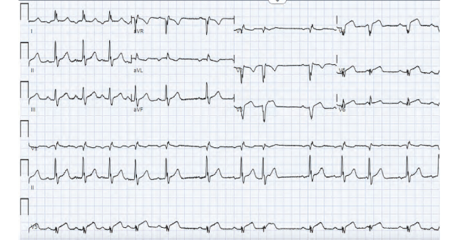
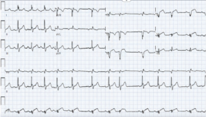
A traditional, left-sided EKG was initially obtained, which demonstrated inverted P waves in lead I, deep Q waves in lead V1, negative QRS complex in V1, and RBBB. It was immediately discerned that the patient had dextrocardia from previous records, and an EKG for dextrocardia was obtained.
The second EKG was concerning for STEMI in the precordial leads (see figure 1). The patient’s first and second troponins from the outside hospital were less than 0.01 ng/mL. The third troponin at our facility resulted as greater than 50.00 ng/mL. The patient was started on IV heparin and immediately taken for cardiac catheterization.
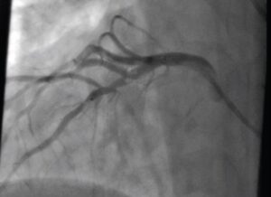
In the cath lab, the patient was found to have evidence of a proximal thrombus and significant stenosis of the LAD (see figure 2). He underwent successful revascularization and stenting of the proximal to mid LAD.
Discussion
Dextrocardia is a rare congenital anomaly where the heart is intrinsically positioned in the right hemithorax with the apex pointing towards the right caudal position.1 It has a prevalence of 0.01 percent.1 Dextrocardia can be associated with an overall situs inversus, where all internal organs are in the reversed position or be limited to situs ambiguous, where only some organs are in the reversed.1
Despite the rarity of dextrocardia, coronary artery disease can occur with a similar frequency to that of the general population.3 Coronary artery disease in a patient with Dextrocardia can present with particular findings on a traditional left-sided EKG that raise suspicion for this anomaly. However, there may be diagnostic dilemmas if these findings are not immediately recognized. This delay in recognition can result in the inadvertent underdiagnosis of STEMIs. Thus, it is important to recognize dextrocardia and adjust our diagnostic tools appropriately.
Dextrocardia with STEMI is a rare clinical presentation that presents with both diagnostic and technical challenges. A literature search through PubMed yielded fewer than 80 case reports of this presentation. Further, patients with dextrocardia may have atypical presentations of STEMIs. Dextrocardia can have features of right axis deviation, positive QRS complexes (upright P and T waves) in aVR, negative P and T waves and QRS complexes in lead I, and absent R wave progression in the precordial leads with dominant S waves.4 In cases of dextrocardia, precordial leads should be placed in a mirror image on the right side of the chest, as is done for a right-sided EKG, with the additional reversal of the right and left limb leads.4
Symptoms of acute coronary syndrome classically present on the left chest wall, however, our patient’s pain was all localized to the right side of the chest, which has been described with other cases of dextrocardia.5 The patient’s initial left-sided EKG did not demonstrate concerning ST segment changes. However, the patient had known dextrocardia based on documented medical history and was confirmed with a recent chest x-ray. Upon the prompt reversal of EKG leads for dextrocardia, the patient was found to have an obvious STEMI in the precordial leads. The patient was then emergently taken for cardiac catheterization. It is important to discern cardiac anomalies, such as dextrocardia, early in a patient’s clinical presentation, as it can significantly impact the timely interpretation of EKGs and the appropriate management of the patient’s care.
 Dr. Ahdi is a senior emergency medicine resident at Corewell Health William Beaumont University Hospital.
 Dr. Barish has been a clinical emergency physician for 37 years and the system professor at William BEAUMONT Oakland University medical school.
References
- Maldjian PD, Saric M. Approach to dextrocardia in adults: review. AJR Am J Roentgenol. 2007;188(6 Suppl):S39-S38.
- Totaro P, Coletti G, Lettieri C, Pepi P, Minzioni G. Coronary artery bypass grafts in a patient with isolated cardiac dextroversion. Ital Heart J. 2001;2(5):394-396.
- Hynes KM, Gau GT, Titus JL. Coronary heart disease in situs inversus totalis. The American Journal of Cardiology. 1973;31(5):666-669.
- Nickson C, Buttner R. Dextrocardia. life in the fast lane. July 19, 2021. Accessed December 1, 2023. Available at: https://litfl.com/dextrocardia-ecg-library.
- Kong B, Wang N, Dou L, Cao D. Coronary angioplasty in an adult with dextrocardia and single coronary artery with the right coronary artery originating from the left anterior descending artery. Coronary Artery Disease. 2019;30(5):390-392.
- Burns E, Buttner R. Right Ventricular Infarction. Life in the Fast Lane. February 8, 2021. Accessed December 1, 2023. Available at: https://litfl.com/right-ventricular-infarction-ecg-library.
Pages: 1 2 3 | Multi-Page


No Responses to “Case Report: A Rare Congenital Heart Anomaly”