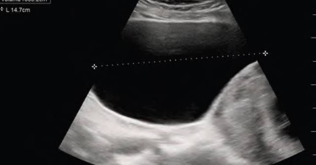
Case
A 40-year-old female at 11 weeks, five days gestation presented to the emergency department (ED) with concern for lower abdominal pressure and inability to fully empty her bladder. She had urinary dribbling when standing and leaning forward. She denied fevers, back pain, saddle anesthesia, numbness, or weakness in the extremities. She reported no history of prior back surgeries, intravenous drug use, recent falls, trauma, or known inciting event. There was no vaginal bleeding or vaginal discharge. Transvaginal ultrasound (TVUS) performed in the outpatient setting a couple of weeks prior to presentation confirmed an intrauterine pregnancy (IUP) with a retroverted uterus. Straight catheterization resulted in approximately 1,500 mL of clear urine output and relief of symptoms. Urinalysis showed no evidence of infection, and the patient was discharged home.
Explore This Issue
ACEP Now: Vol 43 – No 01 – January 2024The patient returned the next day with concern for ongoing urinary retention. She had only been able to void small amounts since the prior visit. Foley catheterization was again performed with approximately 1,200 mL of urine output and relief of symptoms. The obstetrics team was consulted due to the urinary retention. The cervix was not visualized during pelvic examination, but manual examination identified the cervix tucked under the pubic bone in an anterior position, concerning for an incarcerated gravid uterus (IGU). The patient was admitted to the obstetrics service and underwent spinal anesthesia in the operating room to manually reduce the uterus. A pessary was inserted during that procedure. On the next day, the Foley catheter was removed and the patient was able to spontaneously void. She was discharged in stable condition with close follow-up.
Analyzing a Rare Diagnosis
Incarcerated gravid uterus is a rare diagnosis with an estimated incidence of one in 3,000 to one in 10,000 pregnancies.1-3 It most commonly occurs in patients with a retroverted uterus during weeks 10 through 16 of pregnancy. As the uterus grows, it can spontaneously correct; however, if the uterine fundus remains in the pelvis it can become trapped against the sacral promontory while the cervix is trapped against the pubic symphysis.1,4 Approximately 15 to 20 percent of all pregnancies occur in a retroverted uterine position, however only a rare minority of those pregnancies lead to IGU.1,5
Risk factors include prior abdominal surgeries, history of pelvic inflammatory disease, or prior history of uterine incarceration.6 IGU can be identified via history and physical, pelvic examination, or MRI. Alternatively, ultrasound can be utilized as a diagnostic modality. Transabdominal ultrasound is more helpful for IGU diagnosis than TVUS as the transabdominal approach allows for better visualization of the cervix in relation to the uterus.5,7 Since IGU is a rare presentation, the literature primarily consists of case reports and case series. Treatment options include Foley catheterization until the uterus spontaneously reduces and adopts a normal vertex presentation, having the mother adopt a knee-to-chest position, or manual reduction with appropriate pain control. More invasive techniques such as colonoscopic or laparoscopic reduction have been described.1,7
Pages: 1 2 3 | Single Page


No Responses to “Case Report: Acute Urinary Retention in a Pregnant Woman”