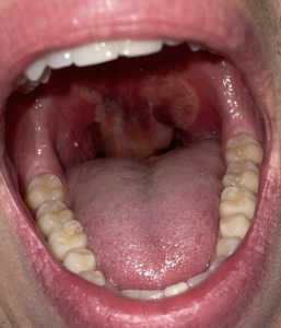
A healthy 20-year-old male presented with one day of a progressively worsening sore throat with difficulty swallowing, increased secretions, and increased work of breathing. He had no history of allergies, anaphylaxis, or recent dental procedures or infection. He endorsed daily inhalational marijuana usage.
Explore This Issue
ACEP Now: Vol 43 – No 11 – November 2024Description of Diagnosis, Management
On examination, he had a significantly enlarged uvula with erythema and edema. His voice sounded muffled. He had mild ecchymosis along the medial soft palate with superficial mucosal irritation. He had no tonsillar edema or exudate. The tongue and lips were normal in size without any swelling. No brawny edema to the submental region. He was tachypneic with increased salivation, but he was able to manage his secretions. The remainder of the exam was unremarkable.
His initial workup consisted of close airway observation with end-tidal capnometry, rapid strep test, throat culture, respiratory pathogens PCR, and a neck X-ray. A bedside nasopharyngoscopy was conducted; it showed crisp tonsillar structures and no laryngeal edema. A diagnosis of uvulitis was given.

Figure 2. Still image captured from nasopharyngoscopy showing no laryngeal edema. (Click to enlarge.)
After this, close airway monitoring was continued and, given the degree of uvular edema and increased secretions, we empirically treated the patient with intravenous (IV) antibiotics (ampicillin-sulbactam), corticosteroids, antihistamines.
The laboratory testing and neck X-ray were unremarkable for any acute infectious etiology. The patient was admitted to the hospital for airway observation. The uvular edema resolved in 24 hours and the patient was discharged on a course of oral corticosteroids and oral antibiotics.
Uvulitis is inflammation of the uvula that is often caused by the spread of an underlying oropharyngeal infection, such as pharyngitis or epiglottitis. Populations such as young children, unvaccinated or undervaccinated patients, and patients with poor dentition are at an increased risk for developing uvulitis. However, isolated uvulitis is highly unusual and has a possible association with inhalational drug use.1

Figure 3. Still image captured from nasopharyngoscopy showing crisp vocal cords and tonsillar fornixes. (Click to enlarge.)
A review of the literature shows cases of isolated uvulitis occurring in the setting of habitual inhalational drug use such as marijuana, fentanyl, or methamphetamine.2 These cases depict isolated enlargement, erythema, and edema of the uvula. In some cases, the clinical presentation worsened, resulting in acute airway obstruction requiring treatment with empiric antibiotics, racemic epinephrine, intravenous (IV) corticosteroids, and antihistamines with close airway observation.
Pages: 1 2 | Single Page


No Responses to “Case Report: Uvular Edema in a Marijuana Smoker”