In Part 1 of our two-part series on marine envenomations, we reviewed some general information on wound care, antibiotic use, antivenom, and explored some species-specific management. In Part 2, we continue to review species-specific management.
Explore This Issue
ACEP Now: Vol 40 – No 08 – August 2021Phylum: Cnidaria
Class: Hydrozoa
Fire Corals (Millepora alcicornis)
Location: Worldwide (excluding Hawaii) in reefs and shallow waters
Appearance: White to yellow-green seaweed-like growths fixed to rocks and coral. They possess tentacles that extend upward and are roughly 2 m in length.
Pathophysiology and Symptoms: Contact with tentacles causes painful, urticarial lesions that may become hemorrhagic and ulcerate. Symptoms usually resolve within 90 minutes, but they can last up to 72 hours with skin hyperpigmentation that can last several weeks. Rarely, patients will present with mild systemic symptoms (eg, nausea, vomiting, myalgias, dyspnea, anxiety, abdominal pain, headaches, etc.).
Management: Pain is best managed with vinegar. Steroid creams and oral antihistamines can be used for mild urticaria. If severe, oral steroids may be warranted.
Class: Anthozoa
Sea Anemones
Location: Worldwide in deep and coastal waters, often attached to coral or rock
Appearance: Anemones vary in appearance. Most are a single polyp with a cylindrical body. Their mouths are surrounded by cnidocyte-containing tentacles.
Pathophysiology and Symptoms: Anemone venom contains multiple enzymes including cytolytic/hemolytic toxins, neurotoxins, cardiotoxins, and protease inhibitors, which cause symptoms ranging from erythema, pruritis, and blisters to fevers, chills, fatigue, myalgias, and syncope. Skin changes can become permanent in the form of hyper-/hypopigmentation and keloid formation.1
Management: Pain is managed with vinegar. Other symptoms are managed with supportive care.
Phylum: Echinodermata
Class: Echinoidea
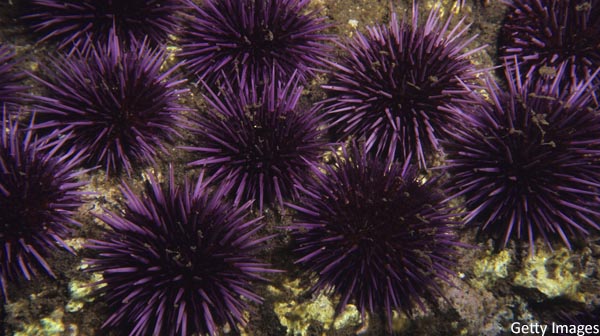
Sea Urchins
Location: Worldwide in both shallow and deep waters
Appearance: Composed of spherical, hard shells called “tests” that measure up to 4–5 inches in diameter which are covered in calcified spines. Venom is contained within these spines, as well as their pedicellarie (ie, pincers), which are more difficult to remove from human skin and contain more venom.
Pathophysiology and Symptoms: Contact causes an erythematous rash with localized burning, pruritis, myalgias (lasting approximately 24 hours), and edema. Symptoms rarely progress to nausea, vomiting, paresthesias, weakness, abdominal pain, hypotension, and syncope. Spines commonly break off, causing hyperpigmentation of the skin, and can lead to granuloma formation, secondary infection, and synovitis, if the joint is involved.
Management: Pain control is generally the biggest concern with these injuries and is best achieved with hot-water immersion and local lidocaine. Attempts to remove spines are often futile. The spines are very fragile and tend to crumble in the skin. To further complicate the removal process, areas where no spine remains may still have the appearance of a foreign body from “tattooing” of the skin. Operative exploration should be considered if there is joint involvement. Granulomas may also need surgical exploration because spines often crumble and are hard to find.
Class: Asteroidea
Crown-of-Thorns Starfish (Acanthaster planci)
Location: Indo-Pacific Ocean, Red Sea, east coast of Africa, and west coast of Central America
Appearance: A central disk with radiating arms (usually more than 15–20 arms), densely covered with spines. Adults are often dull brown to green colored (although some have bright colors to warn predators) and normally range from 9–14 inches in diameter.
Pathophysiology and Symptoms: Spines pierce the skin and cause severe pain (usually lasting ≤3 hours) with local inflammation. The spines are coated with a slime that is extremely toxic and, in severe cases, can cause paralysis, hemolysis, and hepatotoxicity. Additional symptoms include paresthesias, nausea, vomiting, and secondary infection.
Management: Same as Echinoidea/sea urchins (see above).
Phylum: Porifera
Class: Demospongiae
Fire Sponge (Tedania ignis), Poison-Bun Sponge or Touch-Me-Not Sponge (Neofibularia nolitangere), Red Moss or Red Beard Sponge (Clathria prolifera), and Australian Stinging Sponge (Neofibularia mordens)
Location: Worldwide
Pathophysiology and Symptoms: These sponges produce crinotoxins (dermal irritants) that cause a maculopapular rash with local edema, bullae formation, paresthesias, and possible joint swelling that resolves spontaneously in approximately seven days. More severe reactions may cause fevers, chills, fatigue, nausea, and myalgias with delayed immunologic responses that manifest as erythema multiforme or dyshidrotic eczema. Additionally, sponges may also be colonized with Cnidaria species and cause a necrotic skin reaction (ie, sponge divers’ disease).2 Note: Rewetting a dried sponge can cause it to regain its toxicity, even after several years.
Management: Control pain with vinegar. Topical steroids and oral anthistamines are used for mild symptoms, and oral steroids for erythema multiforme or dyshidrotic eczema.2–4
Phylum: Chordata
Subphylum: Vertebrata
Family: Elapidae
Subfamilies: Hydrophiinae and Laticaudinae
Stokes’ Sea Snake (Astoria stokesii), Beaked Sea Snake (Enhydrina schistose), and Yellow-Bellied Sea Snake (Pelamis platurus)
Location: Indo-Pacific Ocean (as far north as San Diego, Calif.), Central and South America
Appearance: Variable appearance. All species are venomous and deliver their venom via a set of small front fangs.
Pathophysiology and Symptoms: Initially, symptoms include a painless or mildly painful bite with local inflammation. However, this can rapidly progress to rhabdomyolysis, hemolysis, cardiac dysrhythmias, renal failure, hepatic failure, seizures, and ascending paralysis with subsequent respiratory failure within minutes to hours. Additional symptoms include cranial nerve abnormalities (eg, dysphasia, dysphagia, ptosis), nausea, and vomiting.
Management: A broad laboratory workup with serial measurements should be undertaken, including complete blood count, chemistry panel, creatine phosphokinase, liver function tests (transaminitis is seen in severe toxicity), and urinalysis. Pressure immobilization (not tourniquets) of an affected extremity should be performed. Supportive care, including IV fluids, and observation for at least eight hours is indicated.5 Multiple antivenoms are available though no evidence suggests any one preferred agent—if you have antivenom, give it. Antivenoms include:
- CSL sea snake antivenom: Administer one to three vials (1,000 units per vial) for any evidence of envenomation (with a 1:10 dilution (1:5 for small children) with 0.9% sodium chloride given via IV over 30 minutes). It is reported that up to seven vials have been safely administered.6
- Terrestrial tiger snake antivenom: Effective for all sea snakes. Administer one vial (3,000 units).
- Thai neuro polyvalent antivenom (NPAV): Effective for beaked sea snake or spine-bellied sea snake.
Class: Chondrichthyes
Order: Myliobatiformes
Suborder: Myliobatoidei
Stingrays
Location: Worldwide
Appearance: Flat and cartilaginous with a stinger containing a retroserrate barb and venom glands located on the ventral aspect of the tail.
Pathophysiology and Symptoms: There are two phases to injury. Phase one is due to traumatic injury from the barb and is characterized by significant pain, usually peaking around 60 minutes post-exposure, but which can persist for up to 48 hours.2 This phase accounts for most of the morbidity and mortality due to hemorrhage, injury to vital organs (as was the case with wildlife expert and television personality Steve Irwin), or subsequent infection. Additional symptoms include nausea, vomiting, diarrhea, muscle cramps, and wound necrosis. Phase two is due to venom release, which causes vasospasm and other significant sequelae, including limb ischemia, cardiotoxicity (eg, dysrhythmias, heart block, non-ST segment elevation myocardial infarction, etc.) seizures, coma, and death.
Management: Pain control is best achieved with hot-water immersion and/or local lidocaine administration. Patients should be brought to the operating room for removal of any barbs in the chest or abdomen. Infusion of prostaglandin E1 has resulted in successful salvage of an ischemic leg, but insufficient data exists to recommend this as routine therapy. There is no antivenom available.2,7
Family: Synanceiidae (*Also classified in the family Scorpaenidae)
Stonefish
Location: Indo-Pacific Ocean
Appearance: Grey, mottled, and often covered with algae that allow for camouflage. These fish possess multiple spines that release venom in response to external pressure.
Pathophysiology and Symptoms: These are the most venomous fishes known, with venom likened to that of a cobra. The venom blocks cardiac calcium channels, increases systemic catecholamine release, simultaneously causing diffuse vasodilation, and increased tissue destruction which propagates uptake of its own venom. Initial effects include rapid onset of severe pain, edema, necrosis, and ulceration. Pain tends to peak at 60 minutes but can persist for several days. Additional symptoms include fatigue, weakness, hyper-/hypotension, syncope, dyspnea, delirium, seizures, and limb paralysis. Severe complications include dysrhythmias, heart failure, heart block, cardiogenic pulmonary edema, hemolysis, and compartment syndrome. Death can occur in as few as six hours from the time of envenomation. Venom remains stable for up to 48 hours after the fish has died, and delayed wound healing for weeks to several months is common.8
Management: Pain control and venom neutralization is achieved with hot-water immersion. Heating the site of a stonefish venom injury to 122 degrees F (50 degrees C) for five minutes prevents wound necrosis and hypotension in animal models.9 Local lidocaine can also be used for pain management. Patients should be observed for 6 to 12 hours.
Antivenom includes Stonefish CSL antivenom. All doses are recommended to be given intramuscular due to an increased risk of anaphylactoid reaction. One vial is equivalent to 2,000 units and neutralizes 20 mg of venom. Give one vial for one to two puncture wounds, two vials for three to four puncture wounds, and three vials for five or more puncture wounds.2
Caveats: Watch closely for signs of necrotizing fasciitis due to a high risk for Vibrio vulnificus co-infection—give antibiotics early and observe for signs of compartment syndrome.10
Family: Scorpaenidae (Scorpionfish)
Lionfish (Pterois volitans and Pterois lunulata)
Location: Indo-Pacific Ocean
Appearance: Similar to stonefish, lionfish possess multiple spines that release venom in response to pressure. Their appearance is variable across 12 species in the Pterois genus, but they generally have alternating brown to orange and white stripes or spots.
Pathophysiology and Symptoms: These fish are common to home aquariums and account for the majority of spiny fish-related calls to poison control centers in the United States.12 Initial symptoms include severe pain that peaks within one to two hours with variable skin changes (eg, erythema versus pallor versus cyanosis). Lesions can progress to hemorrhagic bullae with necrosis. Systemic effects are similar to stonefish (see above).
Management: Management is similar to the approach for stonefish envenomation (see above), with the caveat that there is no antivenom for lionfish.
References
- Abdel-Lateff A, Alarif WM, Asfour HZ, et al. Cytotoxic effects of three new metabolites from Red Sea marine sponge, Petrosia sp. Environ Toxicol Pharmacol. 2014 May;37(3):928–935.
- Nelson LS, Howland M, Lewin NA, et al. Goldfrank’s Toxicologic Emergencies, 11e. New York: McGraw-Hill, 2019.
- Schmidt ME, Abdelbaki YZ, Tu AT. Nephrotoxic action of rattlesnake and sea snake venoms: An electron-microscopic study. J Pathol. 1976 Feb;118(2):75–81.
- Southcott RV, Coulter JR. The effects of the southern Australian marine stinging sponges, Neofibularia mordens and Lissodendoryx sp. Med J Aust. 1971 Oct 30;2(18):895–901.
- Tintinalli JE, Ma O, Yealy DM, et al. Tintinalli’s Emergency Medicine: A Comprehensive Study Guide, 9e. New York, NY: McGraw-Hill, 2020.
- Mercer HP, McGill JJ, Ibrahim RA. Envenomation by sea snake in Queensland. Med J Aust. 1981 Feb 7;1(3):130–132.
- Shiraev TP, Marucci D, McMullin G. Threatened limb from stingray injury. Vascular. 2017 Jun;25(3):326–328.
- McGoldrick J, Marx JA. Marine envenomations; Part 1: Vertebrates. J Emerg Med. 1991 Nov–Dec;9(6):497–502.
- Wiener S. Observations on the venom of the stone fish (Synanceja trachynis). Med J Aust. 1959 May 9;46(19):620–627.
- Tang WM, Fung KK, Cheng VC, et al. Rapidly progressive necrotising fasciitis following a stonefish sting: A report of two cases. J Orthop Surg (Hong Kong). 2006 Apr;14(1):67–70.
Dr. Hauglid is an emergency medicine resident at the University at Buffalo.
Dr. Kiel is assistant professor of emergency medicine and sports medicine at the University of Florida College of Medicine-Jacksonville.
Dr Schmidt is assistant professor of emergency medicine at the University of Florida College of Medicine-Jacksonville.


No Responses to “Emergen-Sea Medicine Part 2: Overview of Marine Envenomations”