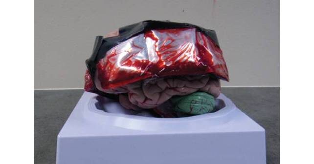
Emergency physicians must be skillful at performing certain high-risk, low-volume procedures. One of these procedures is emergent skull trephination, or burr hole placement, to decompress an expanding epidural hematoma. We are not aware that the procedure is taught routinely in emergency medicine residencies. It is not part of the Emergency Medicine Defined Key Index Procedure Minimums for board certification by the ACGME.1 According to the 2019 Model of the Clinical Practice of Emergency Medicine, intracranial hemorrhage is listed as a “critical” acuity. However, trephination to relieve pressure caused by epidural hemorrhage is not listed as a procedural skill integral to the practice of emergency medicine.2 While the list is not considered comprehensive, there are serious implications of the emergent presentation of this condition that would benefit from widespread training on this skill.
Explore This Issue
ACEP Now: Vol 41 – No 10 – October 2022There is a body of medical literature that suggests substantial outcome benefit when the procedure is appropriately employed. Multiple small studies and case reports indicate improved results in cases of expanding epidural hematomas with early intervention by emergency physicians.3-8
One study in 1996 assessed outcomes associated with traumatic epidural hematomas in 21 patients. The authors found that patients who underwent a drainage craniotomy in less than 70 minutes had a better recovery than those who had one at 90 minutes or more.4 A case described in ACEP Now in 2017 detailed the successful employment of emergency department trephination in a two-year-old male with an excellent outcome.5 Comparable outcomes have been found in similar studies.6-8 If left untreated, the reversal of symptoms becomes more difficult, and complications, including fatality, are positively correlated with increasing degree of uncal herniation.9
Indications
The presentation of epidural hematoma is variable. Classic symptoms that include a lucid interval between two episodes of loss of consciousness are said to occur only 20 percent of the time.10 In general, emergency department trephination should be considered in patients with clinical decline heralded by loss of consciousness—Glasgow Coma Scale less than 8—with or without ipsilateral anisocoria in the presence of a known epidural hematoma, without available neurosurgery. Whenever possible, neurosurgical consultation should take place prior to the initiation of the procedure, and a computed tomography (CT) scan should ideally be obtained prior to initiation of the intervention.11
Procedure Description
On patients, the skull thickness should be assessed utilizing CT and the drill depth set. The procedure site is typically located approximately 2 cm superior and 2 cm anterior to the tragus. A 4-cm vertical skin incision is made. A curved mosquito clamp or periosteal elevator will assist in visualizing the skull. One type of drill that has been described in the literature is the Galt trephine, which has a T handle that is used to create hand pressure coupled with a twisting motion to cut into the skull.3 Figure E shows the instrument we have used. The Galt trephine will create a circular piece of bone that can be removed either with the periosteal elevator or a mosquito clamp. The bone fragment should be placed in saline. At this point, the blood should drain spontaneously. But if needed, small tubing connected to a syringe can be inserted into the wound to aspirate blood.5,12
Pages: 1 2 3 4 | Single Page


No Responses to “Emergency Department Trephination (Burr Hole) for Epidural Hematoma”