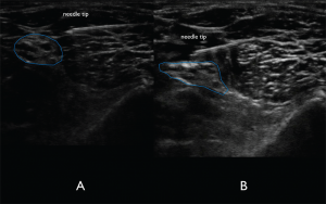
Even though some experienced users may prefer the out-of-plane technique, we feel that the in-plane lateral to medial approach is both easier and safer for the novice sonographer. Slowly advance the spinal needle maintaining a nearly parallel angle to the probe with an in-plane technique (see Figure 6). Since the needle will be entering the lateral thigh, the needle tip will not be visualized immediately (like in other in-plane blocks), and the operator will often have to advance the needle a few centimeters until the tip will be seen entering the scanning plane. Attempt to place the needle tip on the superficial edge of the distal sciatic nerve without penetrating the nerve bundle (see Figure 7A). It is often difficult to determine whether the needle tip is either in the superficial tissue or the fascial plane of the nerve. Injecting small aliquots of either anesthetic or normal saline and visualizing its
Explore This Issue
ACEP Now: Vol 34 – No 06 – June 2015
(Click for larger image)
Figure 7A. In-plane lateral to medial approach of the needle. The needle tip is visualized clearly to ensure lack of nerve and vasculature puncture.Figure 7B. Spread of anechoic local anesthetic surrounding the distal sciatic nerve indicating a successful nerve block. Credit: Arun Nagdev
spread can be the discriminatory test to ensure that the needle tip is in the correct location. Once in the correct location, aspirate to confirm lack of vascular puncture, then slowly inject 2–3 mL aliquots of anesthetic until spread of anechoic fluid is noted to track around the distal sciatic nerve (see Figure 7B). Fanning the probe proximally and distally without moving the needle can be a simple method to confirm tracking of fluid in the fascial plane. We do not recommend novice sonographers place the needle tip on the inferior aspect of the nerve given its relative proximity to the popliteal vasculature. The sciatic nerve in the popliteal fossa has a significant complex fascial sheath. If there is difficulty visualizing anesthetic tracking along the nerve, the operator may need to place the needle tip through the complex fascial sheath, which can resemble epineurium. Often, the operator will feel a “pop” when puncturing the fascia. Using normal saline or very small aliquots of anesthetic under ultrasound visualization will then show a clear demarcation of nerve and fascia.3,4
Complications
Intravascular injection should be a concern because of the proximity of the popliteal vasculature and the distal sciatic nerve. A flat needle angle will allow for clear needle tip visualization and hopefully reduce chances of either local anesthetic systemic toxicity or peripheral nerve injury.
Pages: 1 2 3 4 | Single Page


No Responses to “How to Perform Ultrasound-Guided Distal Sciatic Nerve Block in the Popliteal Fossa”