The Case
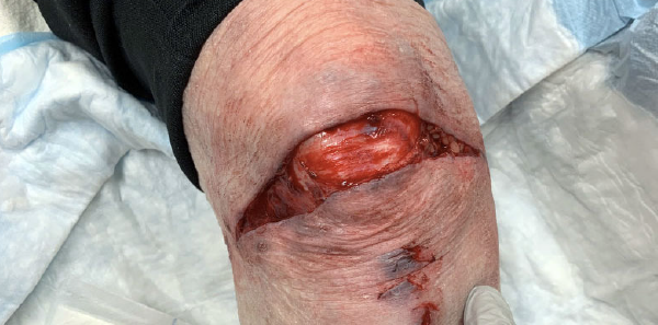
An 81-year-old female presents to the emergency department with a right knee injury after slipping on uneven pavement. She has a large 10-cm laceration that extends across the anterior surface of the knee (see Figure 1). Her patella is exposed; however, there is no clearly visible intra-articular surface to indicate violation of the joint capsule. Radiographs of the knee show no fractures or foreign bodies. How will you determine whether the laceration extends into the joint capsule? Does she need to go to the operating room (OR) for irrigation and debridement (I&D) to prevent septic arthritis?
Explore This Issue
ACEP Now: Vol 39 – No 04 – April 2020Looking for Leaks: The Saline Load Test
First described in 1975, the saline load test (SLT) is the traditional test utilized to assess for traumatic arthrotomy associated with periarticular wounds.1 Using sterile technique, an 18-gauge needle, and syringe, the joint capsule is accessed at an area away from the wound using the same approach for performing arthrocentesis. Intra-articular needle placement is confirmed by aspirating a small amount of synovial fluid. Next, sterile saline is slowly injected until the joint capsule becomes distended. The saline should meet little resistance when injected into the joint capsule, as significant resistance suggests extra-articular infiltration. Extravasation of saline from the periarticular wound indicates the joint capsule has been violated, whereas failure of extravasation indicates an intact joint capsule. After injecting the saline, it is important to wait a few minutes and gently move the joint through its range of motion, as this may make slowly leaking saline more apparent. Finally, aspirate the saline from the joint capsule and withdraw the needle from the patient’s knee.
Adding methylene blue to the sterile saline previously was thought to improve visualization of leakage; however, this practice is not necessary or currently recommended.2 Further, the methylene blue may cause a local inflammatory reaction and interfere with knee arthroscopy should the patient need to go to the OR for I&D.3

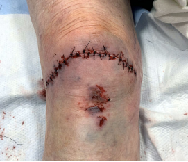
Figure 2: The patient’s knee following wound irrigation and repair.
PHOTOS: Jonathan Strong
How much saline should you inject? Enough to visibly distend the joint capsule or until you meet resistance, indicating the capsule is nearly full. The amount of saline will vary by the size of the joint and the size of the patient, but Roberts and Hedges’ Clinical Procedures in Emergency Medicine and Acute Care generally recommends 100–200 mL for the knee, 40–60 mL for the shoulder, 20–30 mL for the ankle and elbow, and 5 mL for the wrist.3 Fully loading the joint is very important, as an insufficient amount of saline may lead to false-negative test results.
How Well Does SLT Perform?
How well does this test perform in diagnosing traumatic knee arthrotomy? In 1996, Voit et al compared SLT to clinical judgment alone in 50 patients with periarticular traumatic wounds of the knee.4 The study found clinical judgment alone had a sensitivity of 57 percent and a specificity of 61 percent when using SLT as the gold-standard test. SLT changed management in 40 percent of patients, and the authors concluded that SLT is superior to clinical judgment alone. However, the authors did not consider the possibility of false-positive and false-negative SLT results.
Several subsequent studies evaluated the sensitivity of SLT in patients undergoing elective knee arthroscopy.2,5-8 The sensitivity of SLT in these studies varied considerably, ranging from 36 to 99 percent, owing to differences in SLT technique such as the amount of saline injected, the size of the surgical incision, the position of the knee, and whether the knee was moved through its range of motion. Other patient-specific factors, such as age, sex, and body mass index, also may have played a role. Regardless, it is difficult to apply the results of these studies of elective arthroscopic knee surgery patients to patients presenting to the emergency department with traumatic knee injuries.
In 2013, Konda et al used a novel approach to study SLT in 50 patients presenting to the emergency department with periarticular knee wounds.9 In this study, all patients underwent SLT, and those with positive results went to the OR for arthroscopy to confirm the presence or absence of a traumatic knee arthrotomy. Patients with a negative SLT and no other indications for operative management were discharged and monitored for septic arthritis at follow-up. Those who subsequently developed septic arthritis were considered to have a missed traumatic knee arthrotomy. How well did SLT perform in this study? The authors found a sensitivity of 94 percent and a specificity of 91 percent using this approach. Given the prevalence of traumatic knee arthrotomy in this study, the false-positive rate was 16 percent and the false-negative rate was 3 percent. This false-negative rate was attributed to a single patient who had a negative SLT but went to the OR for a grossly contaminated wound and was found to have a traumatic knee arthrotomy. The authors note that none of the patients discharged after a negative SLT went on to develop septic arthritis; however, the study was underpowered to adequately detect infection in this population.
A Loaded Question: Is SLT Obsolete?
In 2013, Konda et al also published a study investigating CT scan as an alternative to SLT.10 In this study of 62 patients presenting to the emergency department with periarticular knee wounds, the presence of intra-articular air on CT scan had 100 percent sensitivity and 100 percent specificity for diagnosing traumatic knee arthrotomy when direct arthroscopic visualization or septic arthritis at follow-up were used as the gold standard for diagnosis. In this same study, the sensitivity of SLT was measured to be 92 percent. The authors conclude that “CT scan performs better than the conventional SLT to identify traumatic knee arthrotomies.” An important caveat is that the study was underpowered to detect patients who go on to develop septic arthritis after a negative CT scan.
This leaves emergency physicians in a difficult spot. Currently, SLT is the generally accepted practice when evaluating a patient for a traumatic knee arthrotomy; however, the evidence behind SLT is weak at best. Should we retire SLT in favor of CT scan? How much evidence is needed to change our practice when the current “standard” practice also has only weak evidence behind it?
This is a difficult philosophical question without a clear answer. However, I can offer some advice for the emergency physician facing such a scenario.
First, discuss the scenario with your fellow emergency physicians and subspecialty colleagues. See if there is a generally accepted practice, policy, or protocol at your institution. It is best not to deviate from the generally accepted practices at your institution unless you have a good reason for doing so. Consider crafting a policy or protocol with your colleagues if your institution does not have one.
Second, discuss the scenario with your patient. They may not understand the nuances of evidence-based medicine, but most will understand uncertainty. Explain your thought process, present the patient with options if you feel it is appropriate, reach a decision together, and document as such.
Finally, consider your own philosophy and values. How much evidence do you need to change your practice? What is your risk tolerance? Are you a traditionalist or an early adopter? Do you place more value in the wisdom of collective past experience or the progress that may come with new innovation? This is for you to decide.
Case Resolution
The case is discussed with the on-call orthopedic surgeon, the patient, and her family. Together, it is decided to proceed with CT scan, which shows no intra-articular air. The patient’s wound is irrigated and repaired, and she is discharged from the emergency department. Follow-up several weeks later reveals she has suffered no infection complications.
Dr. Strong is a clinical fellow in the department of emergency medicine at Brigham and Women’s Hospital in Boston.
References
- Patzakis MJ, Dorr LD, Ivler D, et al. The early management of open joint injuries. A prospective study of one hundred and forty patients. J Bone Joint Surg Am. 1975;57(8):1065-1070.
- Metzger P, Carney J, Kuhn K, Booher K, Mazurek M. Sensitivity of the saline load test with and without methylene blue dye in the diagnosis of artificial traumatic knee arthrotomies. J Orthop Trauma. 2012;26(6):347-9.
- Roberts JR, Custalow CB, Hedges JR, et al. Roberts and Hedges’ Clinical Procedures in Emergency Medicine and Acute Care. 6th ed. Philadelphia, PA: Elsevier; 2013.1092:4.
- Voit GA, Irvine G, Beals RK. Saline load test for penetration of periarticular lacerations. J Bone Joint Surg Br. 1996;78(5):732-733.
- Keese GR, Boody AR, Wongworawat MD, et al. The accuracy of the saline load test in the diagnosis of traumatic knee arthrotomies. J Orthop Trauma. 2007;21(7):442-443.
- Tornetta P, Boes MT, Schepsis AA, et al. How effective is a saline arthrogram for wounds around the knee? Clin Orthop Relat Res. 2008;466(2):432-435.
- Nord RM, Quach T, Walsh M, et al. Detection of traumatic arthrotomy of the knee using the saline solution load test. J Bone Joint Surg Am. 2009;91(1):66-70.
- Haller JM, Beckmann JT, Kapron AL, et al. Detection of a traumatic arthrotomy in the pediatric knee using the saline solution load test. J Bone Joint Surg Am. 2015;97(10):846-849.
- Konda SR, Howard D, Davidovitch RI, et al. The saline load test of the knee redefined: a test to detect traumatic arthrotomies and rule out periarticular wounds not requiring surgical intervention. J Orthop Trauma. 2013;27(9):491-497.
- Konda SR, Davidovitch RI, Egol KA. Computed tomography scan to detect traumatic arthrotomies and identify periarticular wounds not requiring surgical intervention: an improvement over the saline load test. J Orthop Trauma. 2013;27(9):498-504.
Pages: 1 2 3 4 | Multi-Page


One Response to “Saline Load or CT: What’s the Best Test for Traumatic Arthrotomy?”
April 26, 2020
Joel Pasternack, MD, PhD, FACEPGood Article by Dr. Strong.
Clinical judgement can be enhanced as follows: Explore and irrigate wound through full range of motion. Obtain cross table lateral x-ray looking for air under the patella or in supra patella recess, after irrigation. The irrigation process will make the x-ray more sensitive for air without decreasing specificity. Gentle syringe irrigation will force more air into joint than may have entered from original injury.