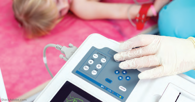
In its simplest form, the solution for tachycardia in children has been the same since the inception of our specialty—find the source. We routinely recognize the source or have a good suspicion of what that source may be. We find the pieces, put together the puzzle, develop differential diagnoses, and risk stratify the potential outcomes. But sometimes it doesn’t all quite make sense. The goal of this discussion is to explore some short topics about pediatric tachycardia for those moments when we think, “Is this kiddo a little too tachycardic?” or “Am I missing something?”
Explore This Issue
ACEP Now: Vol 43 – No 07 – July 2024“How much tachycardia can we attribute to fever?”
After taking into account a child’s agitation and anxiety when recording a heart rate, is there a particular amount of tachycardia that we can expect relative to each degree of fever?
A 2004 prospective study enrolled 490 children younger than one year of age.1 Rectal temperatures were the gold standard, and children who were fussy or crying were excluded. Other known causes of tachycardia were excluded, such as dehydration warranting IV fluid rehydration, hypoxemia, albuterol within four hours, cardiomyopathy, dysrhythmia, sepsis, known endocrine diseases, and anemia. The authors were investigating the relationship between heart rate and temperature increases of 1°C. The mean temperatures in infants with and without fever were 37.2°C and 38.8°C, respectively. In infants younger than two months of age, there was no association between heart rate and temperature. In infants older than two months, the authors found that the mean increase in pulse rate per 1°C temperature increase was 9.6 beats/min (95 percent CI 7.7-11.5 beats/min).
Another retrospective study of 21,033 children evaluated heart rate via the pulse oximeter and temperature via tympanostomy thermometer.2 The authors identified a 10.52 bpm/1°C increase broadly across all ages. Although the increase in heart rate (approximately 10 bpm/1°C rise) is consistent with the earlier study and the data set is large, the data analysis and exclusion criteria are very broad, making this a potentially significant limitation of the latter study’s results. Although earlier studies suggest 10 bpm/1°C, more recent studies may suggest otherwise. A recent retrospective study evaluated 61,321 children with temperatures ranging from 36°C to 40.5°C.3 Children were divided into six pre-determined age groups (zero to less than three months, zero to younger than three months, three months to younger than one year, one to younger than two years, two to younger than five years, five to younger than 10 years, and older than or equal to 10 years). In an effort to exclude any additional factors that may lead to tachycardia, the authors had an extensive exclusion criteria list. Examples included, but were not limited to, any child who required serum labs (including a serum dextrose), children with hypoxia, agitated or crying children, and those with suspected anemia, orthopedic complaints, trauma, environmental factors, overdoses, or acute abdominal complaints that would cause “intense internal pain.” The temperatures were all digital axillary readings rather than rectal or oral temperatures that are typically considered gold standards. For all groups except the zero to three-month age group, the biggest increase in heart rate per 1°C was when the temperature was rising from 37 to 38°C and was about 20 bpm. The range of increase was similar among all age groups when rising from 38 to 39°C and was approximately 10-15 bpm/1°C. The range of variation across all groups from 39 to 40°C was wider at three to 10 bpm/1°C. This study would suggest that children’s heart rates seemed to increase the most just before spiking a fever at 38°C. The 10 bpm/1°C is similar to the prior studies in the 38 to 39°C temperature range but did not seem to hold true in the 39 to 40°C range. A potential limitation of this study’s findings, though, may be the digital axillary temperature readings, which other studies have shown may not be routinely consistent with rectal temperatures.4 A separate 2020 retrospective study found a greater increase in heart rate at 21.5 bpm/1°C in their local subjects and 18.3 bpm/1°C when they analyzed a national database.5 This latter study’s primary goal was to evaluate this same topic in adults, so no exclusion criteria were used, and no summary pediatric data were published.
Conservatively, the best current data suggest an increase in 10 bpm for every 1°C rise in body temperature for fever in children presenting to the ED. Although some studies may suggest larger responses, 10 bpm/1°C is likely the best ballpark number to currently attribute to fever. Although not an ED study, an inpatient pediatric study of 60,863 children found a similar association.6
“Are there any places we may need to consider where children can ‘hide’ disease that may be suggested by tachycardia?”
Although almost any disease can potentially present with tachycardia, two to consider may be pneumonia and myocarditis. In adults, pulmonary embolism (PE) should be considered in cases of persistent or disproportionate tachycardia. Although PE should be considered in children also, it is very uncommon, with a reported rate in the pediatric community of 0.14 to 0.9 cases per 100,000. Anecdotally, pneumonias can sometimes be difficult to diagnose in children. Some lower lobe pneumonias manifest as upper abdominal pain; some simply present as persistent tachycardia. A prospective cohort study of 570 children ages one to 16 years found an odds ratio of 1.3 (95 percent CI; 1.0-1.6) for tachycardia in the setting of a consolidated pneumonia.7 Regarding myocarditis, a separate prospective observational study of 63 cases of pediatric myocarditis found tachycardia (96.8 percent) to be the most common arrhythmia found in these children.8 Although these results don’t suggest that everyone with tachycardia needs a chest radiograph or cardiac evaluation, they do suggest that pneumonia and myocarditis should be on our differential diagnosis when considering the evaluation of children with tachycardia.
“When discharging a child with tachycardia, does having tachycardia suggest that they will bounce back more easily?”
A 2017 study over 44 months retrospectively reviewed children ages two months to 17 years who were discharged from a pediatric ED.9 The authors specifically looked at those patients who discharged with an abnormal heart rate, respiratory rate, temperature, or oxygen saturation level. The primary goal of this study was to evaluate the frequency and nature of significant adverse events within 72 hours in children who had abnormal vital signs at discharge. During this 44-month time period, the pediatric ED discharged 33,185 children—of whom 5,540 (17 percent) had at least one abnormal vital sign. Abnormal vital signs were defined as temperature ≥ 38°C, oxygen saturation < 95 percent, or heart rate and respiratory rate outside published age-specific ranges. Of the 5,540 children discharged with one or more abnormal vital signs, 24 (0.43 percent) had a significant adverse event within the next 72 hours (defined as re-presentation to hospital and admission for greater than or equal to five days, CPR, endotracheal intubation, or unexpected surgery). Death related to the initial visit, even though it may have happened outside the 72-hour window, was explored and included on a case-by-case basis. In the group discharged with at least one abnormal vital sign—compared to the normal vital sign group—the relative risk for a significant adverse event was 2.5 (95 percent CI 1.6-4.2), and the number needed to harm was 380 (95 percent CI 252-767). In the 24 children who were discharged with at least one abnormal vital sign and returned with a significant adverse event, 67 percent demonstrated tachycardia at discharge. In children who were discharged with at least one abnormal vital sign and had no adverse events, 56.1 percent had tachycardia at discharge. Thus, heart rate had poor discrimination for predicting adverse events. There were two deaths in the group with abnormal vitals at discharge; one was an unrelated accidental death and the other was due to infection “not believed to be potentially preventable by hospital observation.” In the group with normal vitals at discharge, there were no deaths. Although discharging a child with abnormal vital signs does appear to increase the risk of bounce back, the likelihood of a significant event is still very low (0.43 percent)—emphasizing the importance of good discharge instructions. Although the most common abnormal vital sign in this study was tachycardia, it was a poor predictor of adverse events.
Similarly, another 2017 retrospective article looked at 126,774 children younger than 19 years of age discharged from two pediatric EDs and four urgent care centers.10 Discharge
tachycardia was defined as greater than or equal to 99 percent for age and was found in 10,470 (8.3 percent) of the patients. Compared to patients who did not have discharge tachycardia, children with discharge tachycardia had an increased risk of revisit within 72 hours of discharge (adjusted RR 1.3; 95 percent CI 1.2-1.5). Although there was an increased risk of bouncing back within 72 hours, children with discharge tachycardia were not associated with clinically important interventions when they returned (RR 1.0; 95 percent CI 0.9-1.0). Clinically important interventions were defined as supplemental oxygen, respiratory medications and admission, antibiotics and admission, and peripheral IV line placement and admission. One child in the cohort bounced back and required CPR, but this child did not have tachycardia at discharge. Again, this study emphasizes the importance of discharge education and instruction.
Pediatric tachycardia can be due to fever or other occult diagnoses, or it may predict an increased risk of return to the ED.
 Dr. Jones is assistant professor of pediatric emergency medicine at the University of Kentucky in Lexington.
 Dr. Cantor is professor of emergency medicine and pediatrics, director of the pediatric emergency department, and medical director of the Central New York Regional Poison Control Center at Upstate Medical University in Syracuse, New York.
References
- Hanna CM, Greenes DS. How much tachycardia in infants can be attributed to fever? Ann Emerg Med. 2004;43(6):699-705.
- Davies P, Maconochie I. The relationship between body temperature, heart rate and respiratory rate in children. Emerg Med J. 2009;26(9):641-643.
- Matsumura O, Morikawa Y, Kaneko T, et al. Relationship between body temperature and heart rate in children with no other apparent cause of tachycardia. Pediatr Emerg Care. 2022;38(12):644-649.
- Craig JV, Lancaster GA, Williamson PR, et al. Temperature measure at the axilla compared with rectum in children and young people: systemic review. BMJ. 2000; 320(7243):1174-1178.
- Kirschen GW, Singer DD, Thode HC Jr, et al. Relationship between body temperature and heart rate in adults and children: a local and national study. Am J Emerg Med. 2020;38(5):929-933.
- Daymont C, Bonafide CP, Brady PW. Heart rates in hospitalized children by age and body temperature. Pediatrics. 2015;135(5):e1173-1181.
- Lynch T, Platt R, Gouin S, et al. Can we predict which children with clinically suspected pneumonia will have the presence of focal infiltrates on chest radiographs? Pediatrics. 2004;113(3 Pt 1):e186-189.
- Rasal G, Deshpande M, Mumtaz Z, et al. Arrhythmia spectrum and outcome in children with myocarditis. Ann Pediatr Cardiol. 2021;14(3):366-371.
- Winter J, Waxman MJ, Waterman G, et al. Pediatric patients discharged from the emergency department with abnormal vital signs. West J Emerg Med. 2017;18(5):878-883.
- Wilson PM, Florin TA, Huang G, et al. Is tachycardia at discharge from the pediatric emergency department a cause for concern? A nonconcurrent cohort study. Ann Emerg Med. 2017;70(3):268-276.e2.
Pages: 1 2 3 4 | Multi-Page


No Responses to “Tips for Tachycardia in Children”